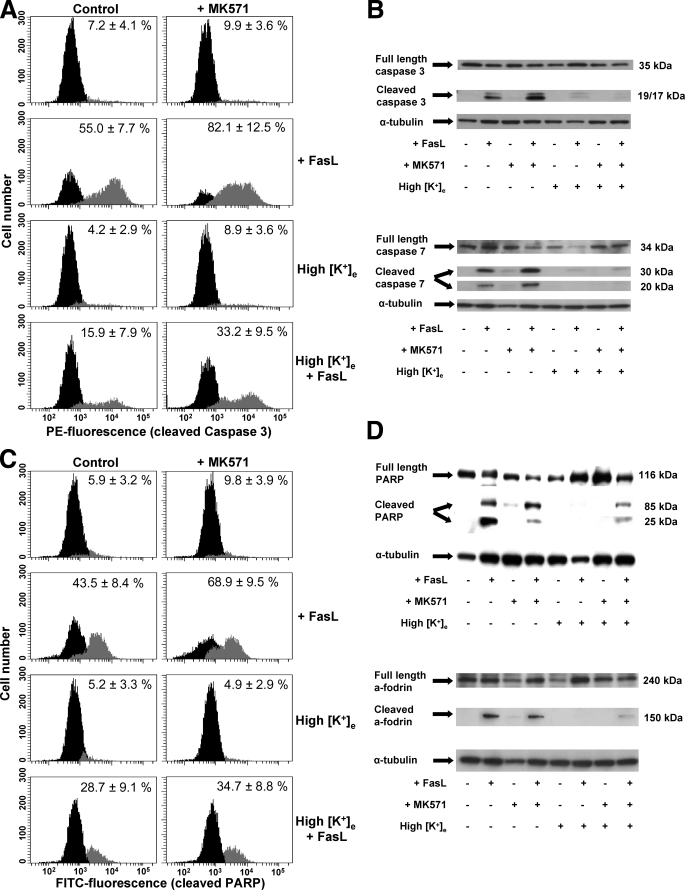
FIGURE 6.
GSH depletion modulates the execution phase of apoptosis by regulation of K+ loss. Apoptosis was induced by 25 ng/ml FasL during 4 h in the presence or absence of 50 μm MK571 and high extracellular K+ medium (High [K+]e). The execution phase of apoptosis was evaluated by the activation of execution caspases (3 and 7) and cleavage of their substrates (PARP and α–fodrin). A and C, immunolabeling detection of cleaved caspase 3 and PARP was performed by single cell analysis using FACS. Frequency histograms in control panels show the distribution of cells with background fluorescence for PE-conjugated anti-active caspase 3 or FITC-conjugated anti-cleaved PARP antibodies (black). A second population with increased fluorescence for PE or FITC (gray) indicates the cells with activated/cleaved caspase 3 (A) and PARP (C), respectively. Plots are representative of at least four independent experiments, and the percentages are means ± S.E. representing the population of cells with active caspase 3 (A) or cleaved PARP (C). B and D, Western immunoblot analysis was done on whole-cell lysates of experimental samples, and blots were incubated with the corresponding antibodies as explained under “Experimental Procedures.” Blots were stripped and reprobed for α-tubulin to verify equal protein loading and are representative of at least three independent experiments.

