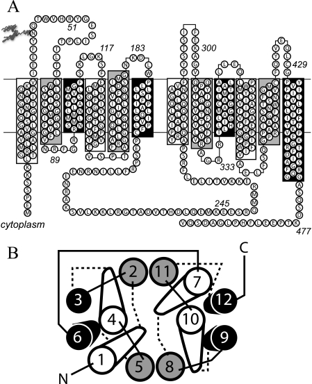
FIGURE 1.
Putative GLUT1 topology and helix packing. A, GLUT1 topology adapted from the GlpT homology model (11). Group 1 TMs are highlighted in white. Group 2 and Group 3 TMs are highlighted in gray and black, respectively. Some TMs extend beyond the bilayer boundaries (indicated by horizontal lines). The bilayer-embedded region of TMs 1–12 comprise amino acids 17–39, 64–86, 93–112, 120–141, 157–178, 187–207, 267–291, 305–325, 335–356, 362–385, 401–421, and 431–452, respectively. GLUT1 is glycosylated at Asn45. TMs 6 and 7 are linked by the large cytoplasmic loop (L6–7). B, putative helix packing arrangement viewed from the cytoplasmic surface. TMs are numbered and colored as in A. Cytoplasmic and exofacial loops are indicated by solid and dashed lines, respectively. The figure is adapted from Refs. 11, 13, and 14.

