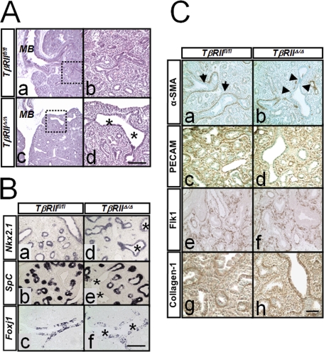
FIGURE 4.
H & E staining and immunohistochemical analysis for lung developmental markers in TβRIIΔ/Δ and control lungs. A, gross histology of lungs from TβRIIfl/fl (panels a and b) and TβRIIΔ/Δ (panels c and d) E15.5 embryos. Saggital sections of lungs were analyzed by H & E staining. MB, mainstem bronchus. Asterisks show dilated proximal airways. Panels b and d are high magnification of areas within dotted squares in panels a and c, respectively. Scale bar, 330 μm for panels a and c; 100 μm for panels b and d. B, cell differentiation in TβRIIΔ/Δ embryonic lungs. In situ hybridization for Nkx2.1 (panels a and d), SpC (panels b and e), and Foxj1 (panels c and f). Asterisks, dilated airways. Scale bar, 100 μm. C, immunohistochemical analysis for α-SMA (panels a and b), PECAM (panels c and d), Flk1 (panels e and f), and Collagen1 (panels g and h) in E15.5 TβRIIfl/fl and TβRIIΔ/Δ lungs. Arrowheads in panel b show reduced expression of α-SMA surrounding the dilated airways of mutant lungs compared with arrows in panel a (control). Scale bar, 100 μm.

