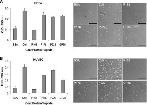
FIGURE 6.
Attachment of human cells to rScl proteins. A, attachment of HDFa. 10 μg of BSA, type I collagen (ColI), or the GP36 synthetic collagen peptide, as well as 100 nm rScl proteins (P163, P176, and P222) were coated onto wells and incubated with HDFa cells. Adherent cells were stained with crystal violet, and the optical density of eluted dye was used to quantify cell attachment to each substrate. Graphic bars depict the average OD recording of triplicate wells from a single representative experiment, and error bars represent the S.D. Digital images of HDFa bound to coated proteins were captured at ×50 magnification prior to elution of crystal violet. The scale bar represents 200 μm. B, attachment of HUVEC. A similar attachment assay was employed as in panel A to assess attachment of HUVECs to rScl proteins and the GP36 synthetic collagen peptide. Graphic bars depict the average OD recording of triplicate wells from a single representative experiment, and error bars represent the S.D. Digital images of HUVECs bound to coated proteins were captured at ×50 magnification prior to elution of crystal violet. The scale bar represents 200 μm.

