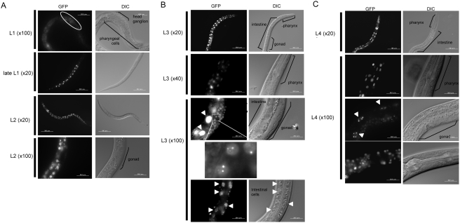
Figure 3. Pqbp-1.1-Venus fusion protein expression during larva stages.
A) At L1 stage, pqbp-1.1-Venus fusion protein is expressed in progenitor neurons in head ganglion. Expression in intestinal cells increases from L1 to late L1 stgae. At L2 stage, pharyngeal and intestinal cells show strong signals. Gonad does not express the fusion protein. B) At L3 stage, pharyngeal and intestinal cells keep a high expression level of pqbp-1.1. Especially, intestinal cells the highest expression level during the development. Somatic gonads also show high signals, and a higher magnification reveals nuclear dots of fusion protein, which is similar to the intranuclear localization of mammalian PQBP1 [11]. Arrowheads indicate intestinal cells. C) At L4 stage, a small number of pharyngeal and intestinal cells show strong signals of pqbp-1.1-Venus. The nuclear dots of the fusion protein are observed in intestinal cells (arrowheads) similarly to L3 stage. Somatic gonads also show weak signals.

