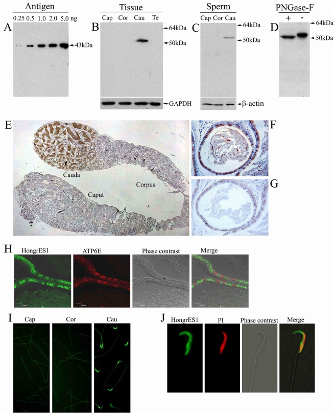
Figure 1. The localization of HongrES1 protein in the rat epididymis.
(A) Rabbit polyclonal antisera were raised against HongrES1 recombinant protein (antigen) and the specificity of the antibody towards HongrES1 was verified by Western blotting. (B) Western analysis of HongrES1 protein in total tissue protein extracts from caput (Cap), corpus (Cor) and cauda (Cau) of the epididymis and testis (Te). Blot was probed with monoclonal antibody against GAPDH to assess protein loading. (C) Western analysis of HongrES1 protein in total protein extracts of sperm from caput (Cap), corpus (Cor) and cauda (Cau) regions of the epididymis. β–actin was used for loading control. (D) The change of molecular masses of HongrES1 protein in total tissue protein of cauda epididymis before (−) and after (+) deglycosylation by Peptide N-Glycosidase F (PNGase-F). (E–G) The immunohistochemical staining showed the expression pattern of HongrES1 protein in the whole epididymis (E) and the epithelial cells of the cauda epididymis (F), meanwhile, the immunoreactivity are also detected in the lumen (E and F). Preimmune serum at the same condition showed background level of immunoreactivity (G). (H) The subcellular localization of HongrES1 protein is determined by using cell-specific antibodies. The immunofluorenscence of HongrES1 (FITC-labeled, green) and ATP6E (Rhodamine labeled for clear cells, red) are shown by confocal microscopy. (I) The localization of HongrES1 protein on the spermatozoa by indirect influorescence assays. The positive HongrES1 immunoreactivity is localized the head of sperm from cauda (Cau) region, but not from caput (Cap) and corpus (Cor) regions of epididymis. (J) The HongrES1 binding pattern on the whole head of the cauda sperm. Sperm DNA was stained with propidium iodide (PI) in red color.

