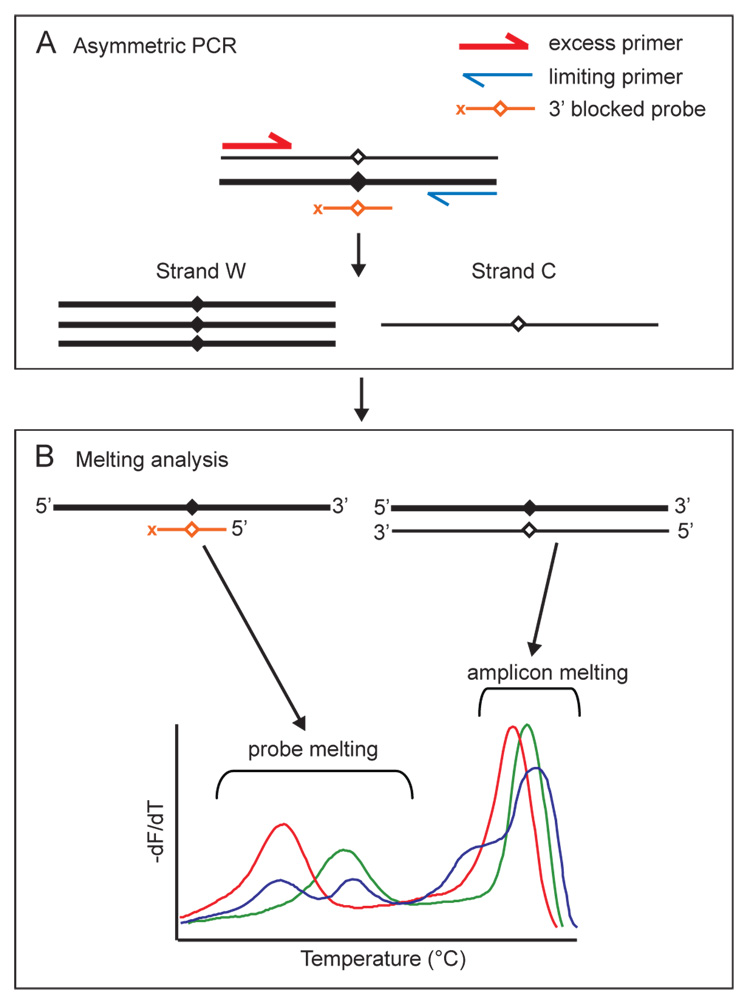
Figure 4.
Combined unlabeled probe and amplicon melting analysis. (A) Asymmetric PCR produces excess copies of strand W and limiting copies of strand C. Excess copies of strand W are available for probe binding, revealing expected and variant sequences under the probe as melting peaks. Sufficient copies of strand C are present so that the full length amplicon duplexes allow variant scanning anywhere within the amplicon. (B) Both product/probe melting transitions and product/product melting transitions are visible on a single negative derivative plot of normalized background corrected fluorescence. Examples of wild-type (green), mutant (red) and heterozygous (blue) melting peaks are illustrated.

