Figure 1.
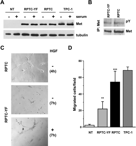
Met expression/activation and morphogenic/motogenic phenotype induced by ectopic expression of RET/PTC1 oncogene in human thyrocytes. (A) Western blot analysis of Met protein expression in thyrocytes uninfected (NT), infected with RET/PTC1 (RPTC), or the RET/PTC1-Y451F mutant (RPTC-YF) and in the RET/PTC1-positive PTC cell line TPC-1. Whole cell lysates were obtained after 24 hours in complete (+) or in serum-free (-) medium. Antitubulin blot is shown as a control for loading. (B) Met tyrosine phosphorylation in RPTC-YF and RPTC cells. Met protein was immunoprecipitated from cells serum-starved for 24 hours, and its phosphorylation was analyzed by Western blot analysis using a pan anti-ptyrosine antibody (pY). The filter was reprobed with anti-Met antibody after stripping. (C) Early tubulogenesis assay. Cells were seeded in Matrigel-coated wells in the absence of serum, with (+) or without (-) HGF. Four hours after plating, extensive tubule networks were formed by RPTC thyrocytes. RPTC-YF cells formed branched cellular cords after 7 hours of incubation only in the presence of HGF. Original magnification, x400. (D) Spontaneous cell migration. Cells were subjected to a migration assay in serum-free medium. Migrating cells were counted under a light microscope and reported as cell number per field. Columns represent mean values ± SD of two independent experiments. **P < .005, ***P < .0005 versus NT.

