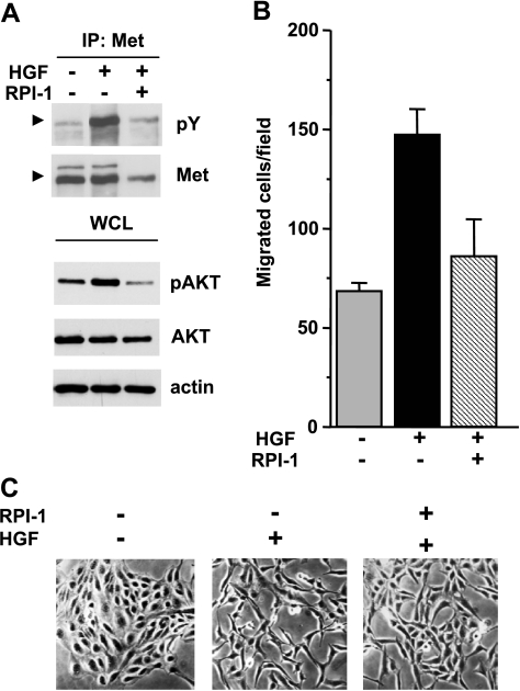
Figure 3.
Biochemical and motogenic effects of HGF in TPC-1 cells. (A) Activation of Met and Akt. Serum-starved cells were treated for 18 hours with solvent (-) or 15 µM RPI-1 (+) and then stimulated with HGF for 10 minutes. Met was immunoprecipitated (IP), and its tyrosine phosphorylation was examined by Western blot analysis using a pan anti-ptyrosine antibody (pY). Arrows indicate the Met mature form. Western blot analysis of Akt activation was performed on whole cell lysates using an antibody recognizing Akt phosphorylated at S473 (pAkt). Filters were stripped and reprobed with anti-Akt and anti-actin antibodies. (B) Cell migration. Cells were exposed to vehicle or to 15 µM RPI-1 for 24 hours and then transferred in Transwell chambers in serum-free medium with or without HGF. Migrated cells are reported as number of cells per field. One experiment representative of three is shown. (C) Cell scattering. Cells were seeded at a low density to allow growing as islets and, 24 hours later, serum-starved and treated with vehicle or 15 µM RPI-1, in the presence or absence of HGF, for 18 hours. Representative images are shown. Original magnification, x100.

