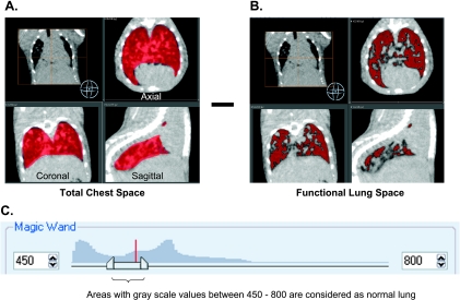
Figure 1.
Method of micro-CT measurement of total tumor burden in lung GEMMs. Tumor and vasculature were measured to represent total tumor burden. The image was adjusted to 450 to 800 grayscale to define boundaries between functional lung area, vasculature, tumor, and chest. (A) Total chest space volume in red includes vessels that come from the heart. (B) Lung area in red with lower CT density was then quantified. Tumor and vasculature were calculated by subtracting the functional lung volume from the total chest space. (C) The total image histogram is shown with the region grow threshold set between 450 and 800.

