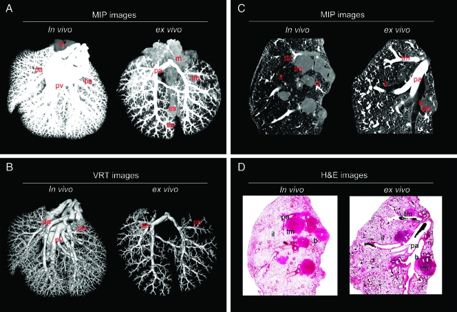
Figure 1.
Micro-CT and histology of an entire murine lung after in vivo or ex vivo injection of Microfil. (A) Three-dimensional MIP images of the entire lung after in vivo or ex vivo Microfil application. (B) Three-dimensional VRT images of the entire lung after in vivo or ex vivo Microfil application. Representative micro-CT and hematoxylin and eosin-stained images clearly showed the course of anatomical structures. (C) Three-dimensional MIP images (in vivo or ex vivo Microfil application) reconstructed from micro-CT images and the corresponding (D) histologic images (in vivo or ex vivo Microfil application). b indicates bronchus; es, esophagus; il, inflated lung; m, mediasternum; pa, pulmonary arteries; pv, pulmonary veins; tm, tumor mass; tr, trachea.

