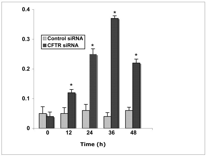
Figure 6.
SULT1E1 activity in HepG2 cells after co-culture in MMNK-1 cell conditioned medium. Lysate was prepared from HepG2 cells maintained in conditioned medium from control- or CFTR-siRNA MMNK-1 cells for 0, 12, 24, 36 or 48 h, then assayed for 20 nM E2 sulfation activity. Results are expressed as pmol E2 sulfated/min/mg protein ± s.d., and represent the means of triplicate SULT1E1 assays in each lysate from triplicate wells. The asterisk represents a significant difference from HepG2 cells maintained in control-siRNA MMNK-1 conditioned medium, at p<0.01 with the Student’s T-test.

