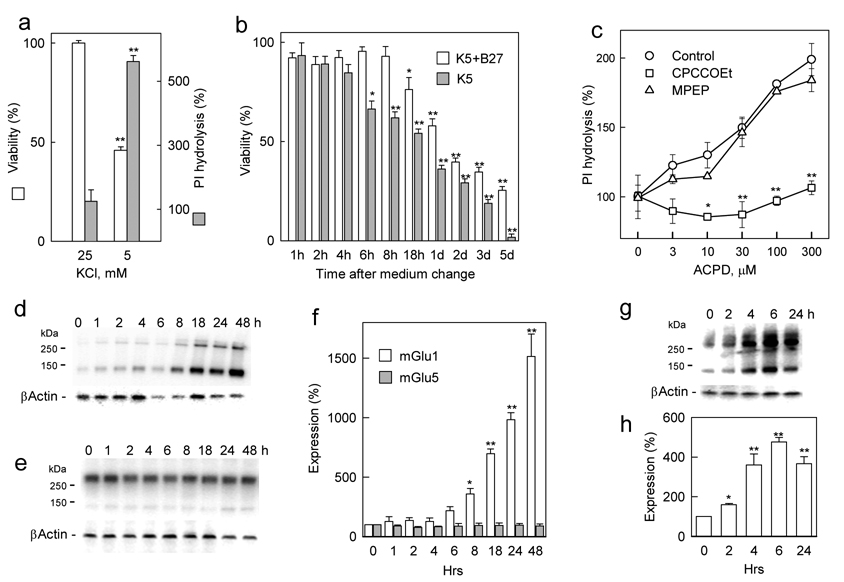
Figure 1. Effect of trophic deprivation on cell viability and mGlu1 expression in primary cultures of rat cerebellar neurons.
a, Cerebellar granule cells were cultured in K25 NB medium supplemented with B27 for one week. At this time, lowering of K+ concentration for 2 days decreased viability and enhanced the expression of group I mGluRs as measured by 100 µM ACPD-stimulated PI hydrolysis (shown as % of basal PI hydrolysis). b, Time course of developing toxicity after medium change from K25+B27 to K5 in the presence and absence of B27. c, The ACPD-stimulated PI hydrolysis, enhanced 24 h after medium change from K25+B27 to K5+B27, is inhibited by the mGlu1 antagonist CPCCOEt (100 µM) but not by the mGlu5 antagonist MPEP (10 µM). Western blots showing that K5+B27 conditions induce a time-dependent increase of mGlu1 (d), but not mGlu5 (e) expression and quantification of these blots after normalization using β-actin (f). Simultaneous removal of B27 accelerates mGlu1 expression as seen on a representative Western blot (g) and quantified after normalization using β-actin (h). All values are means from at least three independent experiments with error bars representing S.E.M. * p<0.05 and **p<0.001 as compared to untreated controls using Student’s t-test.

