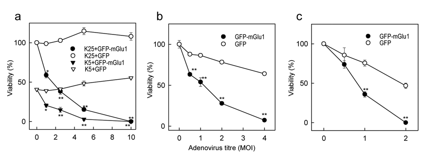
Figure 4. Enhanced mGlu1 expression decreases the survival of neuronal and glial cells.
a, Adenovirus-mediated overexpression of GFP-mGlu1 fusion protein, but not of GFP alone, induces toxicity in rat cerebellar neurons treated with increasing doses (MOI) of adenovirus. Similar toxic effects of GFP-mGlu1 overexpression are seen in mouse cortical neurons (b) and rat cortical astrocytes (c). The cells were infected on the next day after plating. Levels of viral infection were monitored by fluorescence imaging and cell viability was measured by the MTT assay 4 days after viral infections. Data are expressed as per cent of viable cells compared to untreated controls and are means with error bars representing S.E.M. from three separate experiments. *p<0.05 and **p<0.001 as compared to GFP-expressing controls using Student’s t-test.

