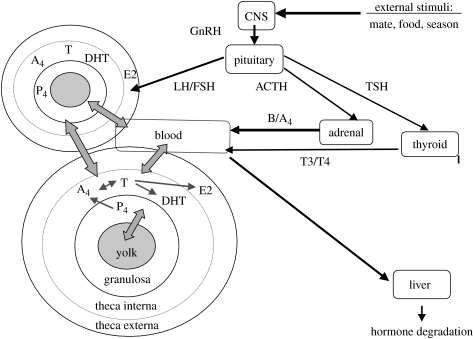
Figure 1.
Diagram depicting hormone production and their accumulation in egg yolk in avian species. The two large circles represent two growing ovarian follicles, including the three steroidogenic layers (theca externa, theca interna and granulosa) in the follicle wall and the oocyte with egg yolk depicted as grey circles. The large rectangle represents a blood vessel and the wide gray arrows possible transport routes for hormones. Large black arrows depict environmental stimuli affecting the female brain (CNS and releasing hormones (e.g. GnRH)), stimulating the pituitary to release hormones (LH, FSH, ACTH and TSH) into the female circulation to regulate the production of peripheral hormones (B, corticosterone; P4, progesterone; A4, androstenedione; T, testosterone, E2, oestradiol; T3/T4, thyroid hormones; DHT, dihydrotestosterone). Small black arrows depict synthesis pathways of ovarian steroid hormones.

