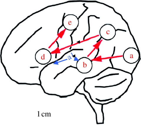
Figure 2.
Schematic of the left hemisphere showing locations and activation sequence for the processing of visual speech (adapted from Nishitani & Hari (2002)). Participants in this MEG study of silent speech-reading identified vowel forms from videoclips. The following regions were activated, in sequence (a) visual cortex, including visual movement regions, (b) superior temporal gyrus (secondary auditory cortex), (c) pSTS and inferior parietal lobule, (d) inferior frontal and (e) premotor cortex. Auditory inputs (primary auditory cortex, A1) are hypothesized to access this system at (b).

