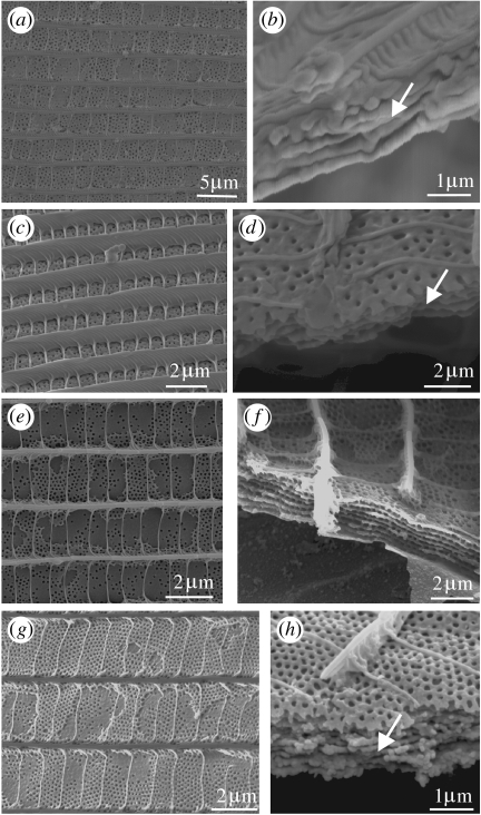
Figure 10.
(a–h) SEMs showing the plan and transverse views of multilayered structures within the lumen. Dorsal-most layer is perforated: subsequent layers are separated by nodules. (a,b) Violet region of Vaga blackburnii (Polyommatinae). (c,d) Blue region of A. o. hollandi (Theclinae). (e,f) Blue region of A. meander (Theclinae). (g,h) Green/blue region of A. chamaeleona (Theclinae).

