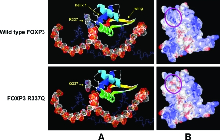
Figure 3.
Predicted structures of the DBD of normal and R337Q FOXP3. A: Predicted interaction of FOXP3 with DNA. FOXP3 is represented in ribbon form, colored blue (NH2-terminal) to red (COOH-terminal), except for residue 337 (labeled) that shows the van der Waals radii of backbone and side chain atoms as dotted surfaces (white, carbon; blue, nitrogen; red, oxygen). Other features referred to in the text (helix 1 and the wing region) are labeled; the main DNA recognition helix (helix 3, green) lies in the major groove of the DNA. The DNA strand predicted to contact FOXP3 R337 is represented as a space-filling model, showing the van der Waals radii of the backbone atoms (white, carbon; red, oxygen; yellow, phosphorus). For clarity, each of the second DNA strands is shown only as a stick model of the backbone (blue). B: Molecular surface of FOXP3, showing areas of positive (blue) or negative (red) electrostatic potential. The structure shown in A has been rotated upward to look toward the DNA-binding surface; the position of residue 337 is indicated by the purple oval, and the DNA strands have been omitted for clarity. Structures were visualized and molecular surfaces were calculated using DeepView (A) or MDL Chime (B) programs.

