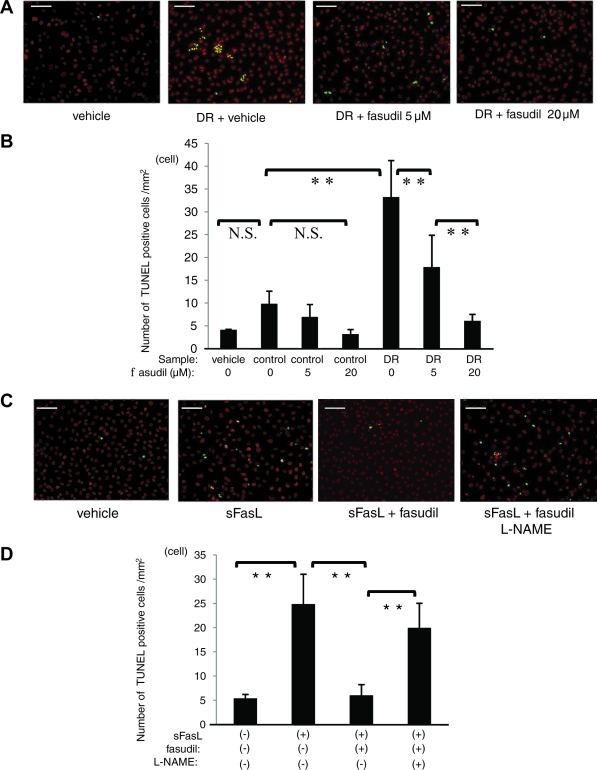
FIG. 7.
Prevention of neutrophil-induced endothelial apoptosis by fasudil. A and B: After pretreatment with 0, 5, or 20 μmol/l fasudil for 1 h, HMVECs were stimulated for 12 h with 10 ng/ml rhTNF-α. Subsequently, unlabeled neutrophils (5 × 10⁵cells/ml) were cocultured with HMVECs for 12 h. Scale bar = 100 μm. The number of apoptotic cells (yellow fluorescence) in four different areas per well was counted (**P < 0.01, NS; n = 15 each). C and D: Involvement of fasudil in sFasL-induced apoptosis was investigated. HMVECs were preincubated with or without 20 μmol/l fasudil before sFasL treatment for 1 h. Furthermore, HMVECs were incubated with or without 1 mmol/l l-NAME before fasudil treatment for 1 h. Scale bar = 100 μm. The number of TUNEL-positive cells in four different areas per well was counted and averaged (**P < 0.01, NS; n = 4 each). (Please see http://dx.doi.org/10.2337/db08-0762 for a high-quality digital representation of this figure.)

