FIG. 1.
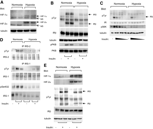
Hypoxia inhibited insulin-induced insulin receptor tyrosine phosphorylation in human and 3T3-L1 adipocytes. A: 3T3-L1 adipocytes were incubated for 16 h at 37°C in normoxia (21% O2) or hypoxia (1% O2). Cell lysates were analyzed by immunoblots with the indicated antibodies. B: 3T3-L1 adipocytes were incubated for 16 h at 37°C in normoxia (21% O2) or hypoxia (1% O2) before being stimulated with insulin (100 nmol/l) for 5 min. C: 3T3-L1 adipocytes were incubated for 16 h at 37°C in normoxia (21% O2) or hypoxia (1% O2) before being stimulated with decreasing concentrations of insulin ranging from 100 to 0.01 nmol/l. D: Cell lysates were subjected to immunoprecipitation using antibodies to IRS-1 or -2 followed by immunoblots using indicated antibodies. E: Human adipocytes were obtained after differentiation of preadipocytes and were incubated for 24 h at 37°C in normoxia (21% O2) or hypoxia (1% O2). After insulin stimulation (100 nmol/l) for 5 min, cell lysates were analyzed by immunoblots with indicated antibodies. Representative experiments of at least three independent experiments performed in duplicate or triplicate are shown. IP, immunoprecipitation; IR, insulin receptor; pTyr, phosphorylated tyrosine; pS6K, phospho-S6K 1; pSer632, phospho-Ser632.

