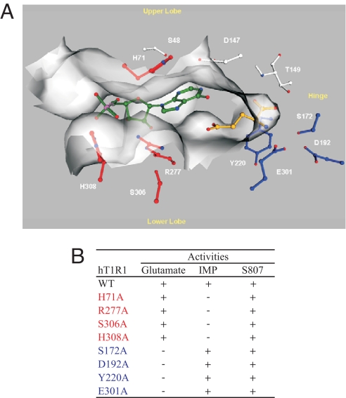
Fig. 4.
A molecular model of T1R1 VFT domain. (A) Key residues for L-glutamate (blue) and for IMP (red) binding. The VFT domain is oriented so that the opening to solvent is horizontal, with the upper lobe on the top and the lower lobe on the bottom. The flytrap hinge region is to the right, and the flytrap opening is to the left. L-glutamate (golden) is located deep inside the VFT domain near the hinge region, while IMP (green) is located close to the opening of the VFT domain. (B) A summary of the mutagenesis data of the key residues. Residues important for L-glutamate activity are in blue, while those for IMP are in red.

