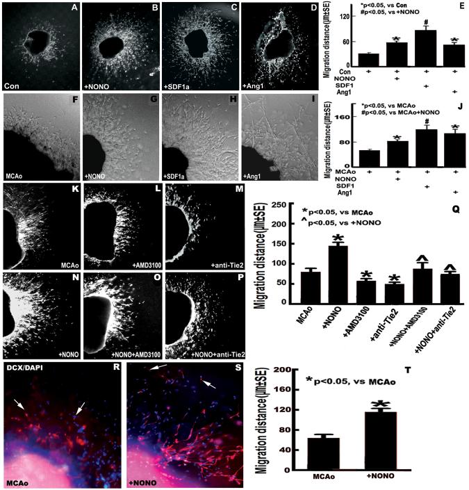
Fig.4.
DETA-NONOate increases SVZ explant cell migration by upregulating SDF1/CXCR4 and Ang1/Tie2 pathways. A-D and F-I: images of SVZ explant cell migration derived from sham and MCAo mice (A and F: control; B and G: + DETA-NONOate 0.4 mg/kg; C and H: + SDF1α 200 μg/L; D and I: + Ang1 200 μg/L). E and J: Quantitative data of SVZ explant cell migration derived from sham (E) and MCAo (J) mice. K-P: images of SVZ explant cell migration derived from MCAo mice (K: MCAo alone; L: + AMD3100 20 μmol/L; M: + anti-Tie2 2 mg/L; N: + DETA-NONOate 0.1 μmol/L; O: + DETA-NONOate 0.1 μmol/L + AMD3100 20 μmol/L; P: + DETA-NONOate 0.1 μmol/L + anti-Tie2 2 mg/L). Q: Quantitative evaluation of SVZ explant cell migration derived from MCAo mice. R and S: DCX-immunofluorecence staining in the cultured SVZ explants derived from MCAo alone (R) and DETA-NONOate treatment (0.4 mg/kg) (S). T: Quantitative data of DCX-positive cell migration. n = 6 well / group.

