Figure 1.
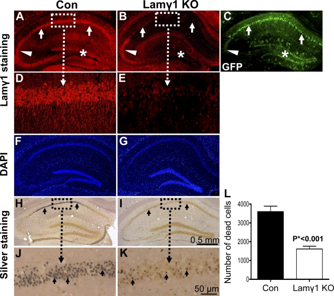
Lamγ1 KO mice were resistant to KA-induced neuronal death in the hippocampus. (A and B) Lamγ1 was expressed in the hippocampal neuronal layers CA1 (A, arrows), CA2/3 (A, arrowhead), and DG (A, asterisk) of control (Con) mice (floxed lamγ1 mice) but was dramatically decreased in the CA1 and DG regions of the lamγ1 KO mice (B, arrows and asterisk). In the CA2 region, lamγ1 was still expressed (A and B, arrowheads). Higher magnification of boxed areas in A and B are shown in D and E, respectively. (C) In the lacZ/EGFP reporter mice that also carry the CaMKII-Cre transgene, GFP (indication of Cre activity) was expressed in the hippocampal neuronal layers CA1 (arrows) and DG (asterisk), whereas GFP was not expressed in the CA2 region (arrowhead indicates background GFP activity). The GFP expression regions correlated well with the regions of lamγ1 disruption in the KO mice. (F and G) DAPI staining revealed a similar pattern of hippocampal neuronal layers between control and lamγ1 KO mice. (H and I) Silver staining shows that intrahippocampal KA injection–induced neuronal death in the CA1 region of lamγ1 KO mice (I) was much less than that of controls (H; H–K, arrows). Higher magnification of boxed areas in H and I are shown in J and K, respectively. (L) Quantitative analysis of KA-induced neuronal death in control and KO mice (seven mice in each genotype). Error bars indicate SEM. Bars: (A–C and F–I) 0.5 mm; (D, E, J, and K) 50 μm.

