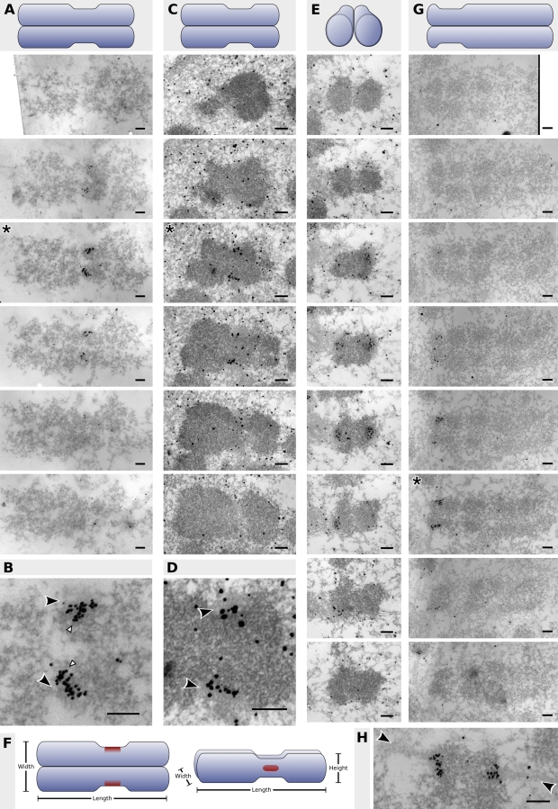
Figure 2.
Localization of CENP-A to the inner kinetochore of human chromosomes. Each panel represents a complete set of serial sections through the centromere, and a schematic of the orientation of each chromosome precedes each set. (A) 60-nm serial sections through an acetone-fixed, colcemid-blocked mitotic chromosome. (C and E) 70-nm serial sections through PFA-fixed, colcemid-blocked mitotic chromosomes in different orientations. (G) 50-nm serial sections through an acetone-fixed unblocked mitotic chromosome. Details of the centromere regions (asterisks) from A, C, and G are shown in B, D, and H, respectively. Black arrowheads in B and D point to the kinetochore outer plate; white arrowheads demarcate putative chromatin fibers illustrated in Fig. 4. Arrowheads in H point to microtubules attached to the kinetochore. (F) A stylized representation of the CENP-A binding domain (red) on human chromosomes drawn to the proportions observed in this study (Table I). Bars, 200 nm.

