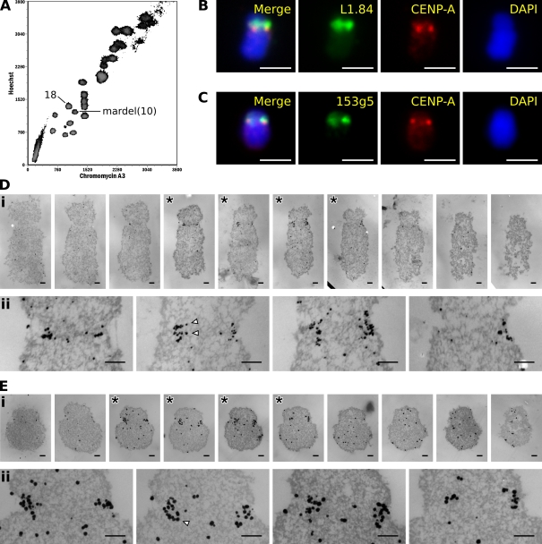
Figure 3.
Localization of CENP-A to the centromeres of flow-sorted chromosomes. (A) Flow-karyotype profile of chromosomes from a human 14ZBHT cell line with the chromosome 18 and mardel(10) populations indicated. (B) Immuno-FISH example of a sorted chromosome 18 using a chromosome 18–specific alphoid probe L1.84. (C) Immuno-FISH example of a sorted mardel(10) chromosome using a bacterial artificial chromosome probe 153g5 specific for the neocentromere. (D) 45-nm serial sections through chromosome 18 (i) and detail of sections exhibiting labeling (asterisks; ii). (E) 45-nm serial sections through a mardel(10) chromosome (i) and detail of sections exhibiting labeling (asterisks; ii). Arrowheads demarcate putative chromatin fibers illustrated in Fig. 4. Bars: (B and C) 2 μm; (D and E) 200 nm.

