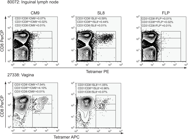
Figure 5. Tetramer+CD3+CD8low/− stained cells from disaggregated lymph nodes and vagina analyzed by flow cytometry.
Disaggregated cells from SIV-infected macaques were stained with Mamu-A*01 Gag CM9, Mamu-A*01 Tat SL8, and negative control Mamu-A*01 FLP tetramers; counterstained with CD3 and CD8 antibodies and analyzed by flow cytometry. Populations of SIV-specific tetramer+CD3+CD8low/− lymphocytes were not detected above negative control staining. However, populations of tetramer+CD3+CD8low cells were detected above background levels. Representative data from inguinal lymph node from animal #R80072, and vaginal submucosa from #27338 is shown. Note negative control staining FLP was not done with the vaginal submucosal cells.

