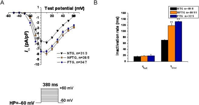
Figure 2. Electrophysiological effects of Cav1.2α1C overexpression in mouse ventricular myocytes.
(A) Averaged peak current-voltage relationships demonstrate significant increase of I Ca,L density at multiple depolarizing pulses in NFTG and FTG compared with NTG cardiomyocytes. The voltage protocol used to record I Ca,L is shown in the inset. (B) Inactivation time constants (τfast and τslow) were determined from I Ca,L traces depolarized to +10 mV fitted by double exponential equation: Y = Ymin+ A1×[1−exp(−t/τfast)] + A2×[1−exp(−t/τslow)], where Y is the fraction of recovery, A1 and A2 are the maximum values of the fast and slow component, and τfast and τslow are the time constants, respectively.

