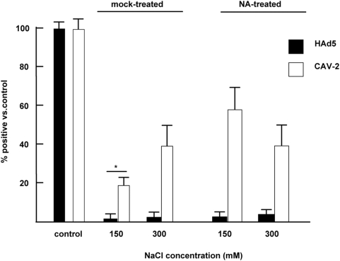
Figure 4. HAd5 and CAV-2 binding to human erythrocyte.
CAVGFP or AdGFP was incubated with 2.5×10⁷ erythrocytes at 1 particle per cell (equivalent to ∼5×10⁹ pp in 1 ml of blood) in PBS and ± additional NaCl. The erythrocytes were pelleted by slow speed centrifugation, and an aliquot of the supernatant was removed and incubated with a monolayer of the most permissive and sensitive cells (911 cells for AdGFP and DKCre cells for CAVGFP). These latter cells were analyzed for GFP expression by flow cytometry 24 hr post-incubation. The percent of GFP+ cells versus the control is shown for mock- or neuraminidase-treated erythrocytes. All samples are significantly (P<0.05) different from the controls. In addition, the star (*) corresponds to a P value of <0.01 between the mock-treated erythrocytes at physiologically relevant salt concentration (150 mM) and the controls.

