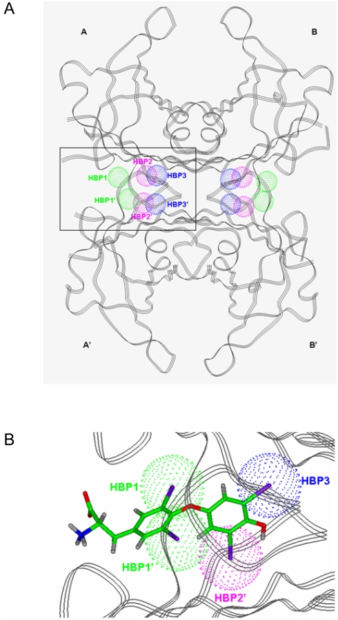
Figure 1.
A) Ribbon diagram of the quaternary structure of TTR with a schematic representation of the three-related pairs of pockets capable of accommodate an iodine atom in each binding site located at the interface of monomers A–A′ and B–B′. These pockets are named in the literature HBP1-HBP1′ (green spheres), HBP2-HBP2′ (pink spheres) and HBP3-HBP3′ (blue spheres). B) Detailed view of one of the binding sites for the TTR:T4 complex, showing the occupation of four of the six HBPs by the iodine atoms of T4 .

