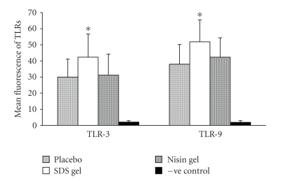
Figure 2.
Flow cytometric analysis of TLR expression in rabbit CVE cells after intravaginal administration of placebo, nisin (15.15 mM in 2 mL of 1% polycarbophil gel) SDS (56 μM in 2 mL of 1% polycarbophil gel) and 2 mL of 1% polycarbophil gel. Mean fluorescence intensity for TLR-3 and 9 were significantly increased in SDS gel-treated animals compared to their respective placebo and nisin gel-treated groups. The mean and error bars indicate standard deviation of triplicate measurements obtained from three separate experiments.

