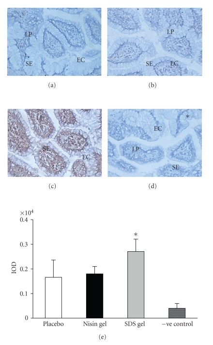
Figure 4.
Immunohistochemical localization of IL-4 in the CVE after intravaginal application of nisin (15.15 mM in 2 mL of 1% polycarbophil gel) (b), SDS (56 μM in 2 mL of 1% polycarbophil gel) (c) and 2 mL of 1% polycarbophil gel (a) for 14 consecutive days. To rule out the nonspecific binding, sections were incubated in preimmune sera instead of primary antibody and considered as negative control (d). Semi-quantitative comparison by measuring integrated optical density (IOD) of immunoreactive IL-4 in treated and placebo is shown (e). The IOD in each area was calculated for the brown color immunoprecipitates. Each IOD value measured is the mean ± SD of six observations obtained from three independent samples. (Mag X 20) (EC = epithelial cells, SE = Squamous epithelium, LP = lamina propria), (* = value is statistically significant at P < .05).

