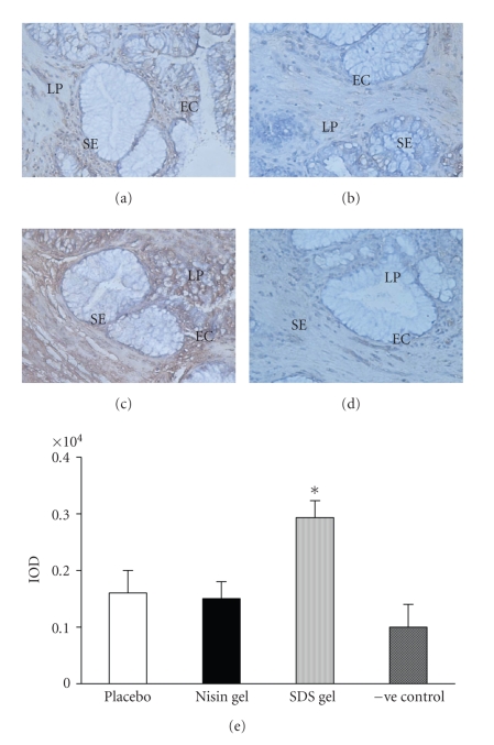
Figure 7.
Immunohistochemical localization of TNF-α in the cervicovaginal tissue after intravaginal application of nisin (15.15 mM in 2 mL of 1% polycarbophil gel) (b), SDS (56 μM in 2 mL of 1% polycarbophil gel) (c) and 2 mL of 1% polycarbophil gel (a) for 14 consecutive days. To rule out the nonspecific binding, sections were incubated in preimmune sera instead of primary antibody and considered as negative control (d). Semiquantitative comparison by measuring integrated optical density (IOD) of immunoreactive TNF-α in treated and placebo is shown (e). The IOD in each area was then measured for the brown color immunoprecipitates. Each value is the mean ± SD of six observations obtained from three independent samples. Figure shown is representative picture from three independent experiments. (Mag X 20) (EC = epithelial cells, SE = squamous epithelium, LP = lamina propria) (* = values are statistically significant at P < 0.05).

