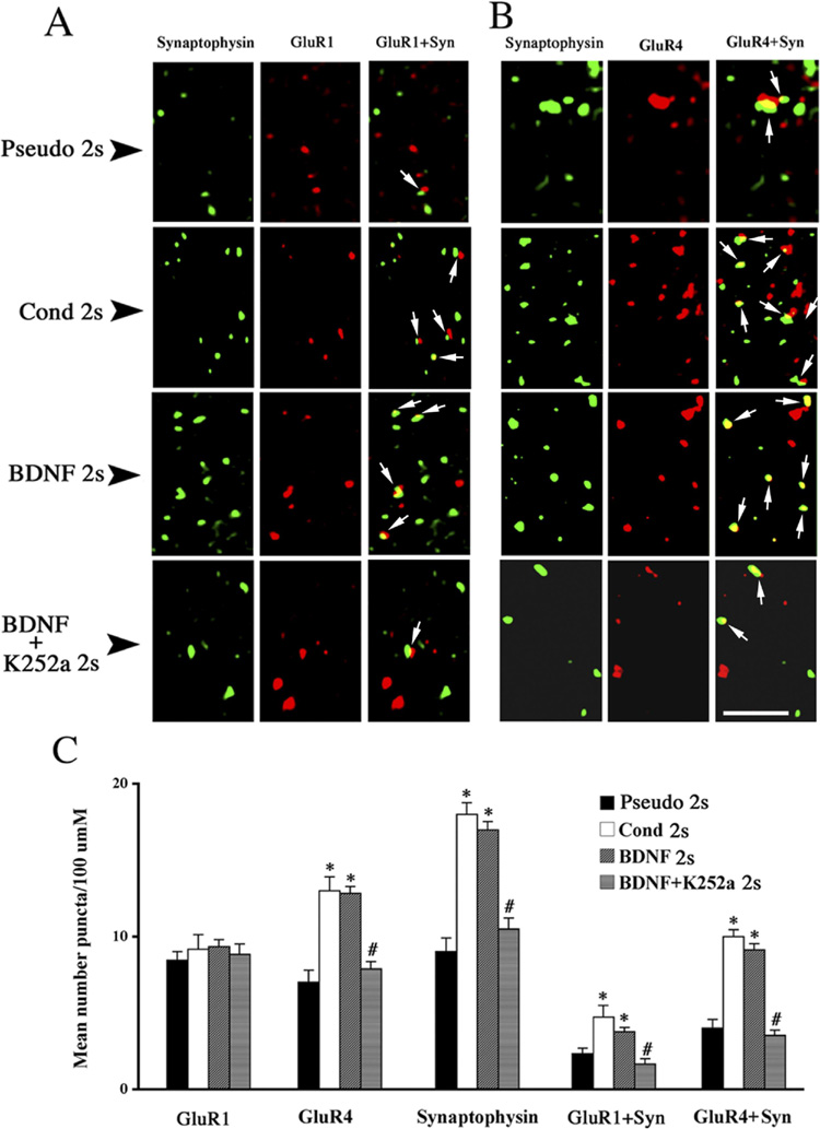
Fig. 7. Localization studies using confocal microscopy reveal that BDNF mimics enhanced synaptic delivery of GluR1- and GluR4-containing AMPAs observed during early stages of conditioning.
(A) Confocal images of abducens motor neurons showing punctate staining for GluR1 subunits (red) and synaptophysin (green) from each of the four groups examined: pseudoconditioned (Pseudo 2s), conditioned (Cond 2s), BDNF-treated (BDNF 2s), and BDNF with K252a-treated (BDNF + K252a 2s) preparations. (B) Confocal images abducens motor neurons showing punctate staining for GluR4 subunits (red) and synaptophysin (green) from similarly treated preparations as shown in A. In A and B, arrows indicate colocalization of AMPAR subunits with synaptophysin. (C) Quantitative analysis of GluR1, GluR4, synaptophysin, GluR1 + synaptophysin, and GluR4 + synaptophysin staining from the different treatment groups. * indicates significant differences between Pseudo 2s and Cond 2s or BDNF 2s groups; # indicates significant difference between BDNF 2s and BDNF + K252a 2s groups. Scale bar = 2 µm.

