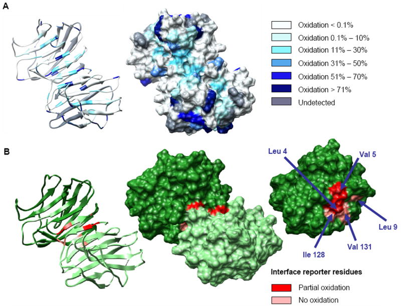
Figure 4.
Per-residue oxidation levels plotted on the solvent accessible surface of dimeric galectin-1 (A). Galectin-1 dimer interface in detail. Reporter residues are indicated in shades of red according to the measured level of side chain oxidation. From left: ribbon structure of the homodimer, solvent accessible surface structure of the homodimer, and monomeric subunit illustrating interfacial residues (B).

