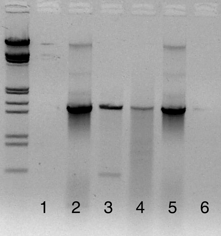
FIG. 2.

Lanes 1, 2, and 3 were amplified using 63F/1542R. Lane 1 is the negative control, lane 2 is the positive control containing only bacterial DNA, and lane 3 is the coral tissue sample. Two bands can be seen in lane 3: the bacterial band at 1.5 kbp and the coral band at 0.6 kbp. These bands are distinctly separated, allowing for the isolation of bacterial DNA. Lanes 4, 5, and 6 were amplified using 8F/1492R. Lane 4 is amplified coral tissue, indistinguishable from the positive control in lane 5. Lane 6 is the negative control. The samples were run on a 1% agarose gel for 1.5 h, with lambda ladders. The gel image has been reversed (i.e., converted to a photo negative) to more clearly show the faint band in lane 3.
