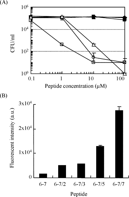
FIG. 4.
(A) CFU of B. subtilis incubated on agar culture medium after the addition of modified peptide under the different concentrations. The modified peptides were added to approximately 105 CFU/ml of cells. After incubation for 1 h at 37°C, the samples were spread on an agar plate and incubated overnight at 37°C. •, peptide 6-7 (original); ▪, peptide 6-7/2; ▴, peptide 6-7/3; ○, peptide 6-7/5; □, peptide 6-7/7; ▵, temporin L (positive control). (B) Fluorescence intensity of FITC-labeled peptides bound onto BacMPs. FITC-labeled peptides (final concentration, 30 μM) were mixed with BacMPs (350 μg/ml) in HEPES buffer for 30 min at room temperature with pulsed sonication. After washing the FITC-labeled peptides five times with HEPES containing 1 M NaCl, the fluorescence intensity of FITC-labeled peptide on BacMPs was measured using a spectrofluorometer (excitation, 495 nm; emission, 520 nm).

