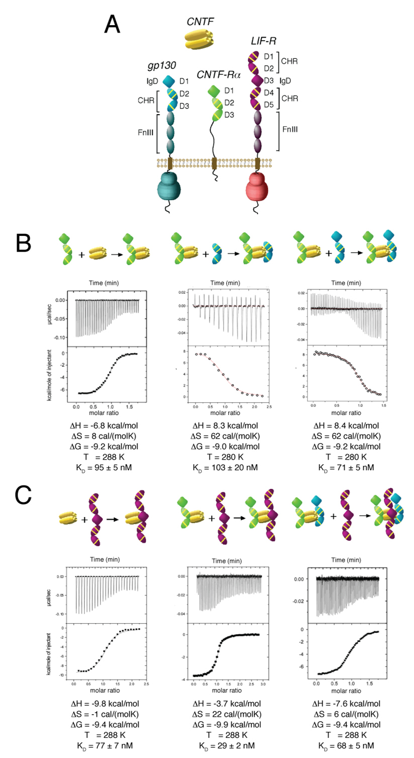
Figure 1. Thermodynamic analysis of the assembly of the gp130/LIF-R/CNTF-Rα/CNTF complex.
(A) Schematic representation of gp130, LIF-R, CNTF-Rα, and CNTF. IgD denotes “Ig-like domain”, while CHR denotes “cytokine-binding homology region” and FnIII denotes “Fibronectin-type III domain”. Each CHR contains three yellow lines to represent conserved disulfides and a WSXWS motif. (B) Isothermal titration calorimetry illustrating the assembly of CNTF and CNTF-Rα, followed by assembly of the CNTF-Rα/CNTF binary complex with gp130 minus or plus the D1 IgD. (C) Titration of LIF-R with CNTF, the CNTF-Rα/CNTF binary complex, and the gp130/CNTF-Rα/CNTF ternary complex. Inset tables list the thermodynamic parameters for the representative binding isotherms.

