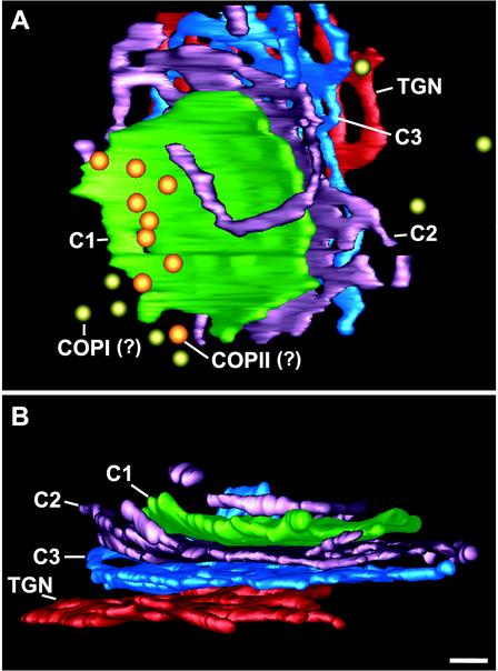
Figure 8.
3D model of a P. pastoris Golgi stack and associated vesicles. A and B are two different views of the same Golgi stack. The model represents the Golgi stack shown in Figure 5 and was generated by rendering a surface fit to the traced contours. This stack has four cisternae. An interesting feature is the tubule from the margin of C2 that reaches around the C1 cisterna. Bar, 100 nm.

