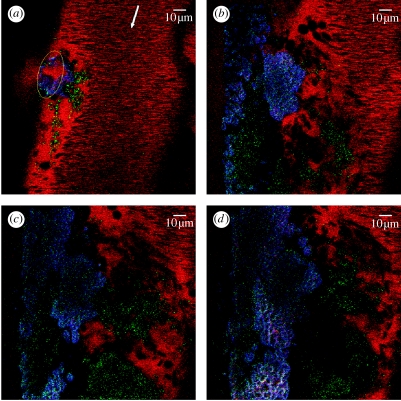
Figure 2.
Intravital confocal images of a developing venous thrombus. (a) Initial state of the thrombus immediately after laser-induced injury (time 0 s). (b–d) Status of thrombus at t=296, 480 and 746 s, respectively. During this time, the number of activated platelets incorporated in the thrombus increases, a fibrin network develops on the injured vessel wall covering the observed length (146 μm) and blood cells are incorporated into the growing thrombus. Not evident from the still frames, the growing thrombus induces turbulent flow around the thrombus that affects thrombus development. At the end of the recording period, the thrombus extended 18 μm from the vessel wall (calculated from Z stack; data not included).

