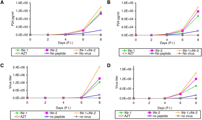
Figure 7. Stimulation of HIV-1 replication by the INrs.
The INrs (12.5 µM) were incubated with H9 cells (A and C) and Sup-T1 lymphocyte cells (B and D) which were then infected with HIV-1 (MOI 0.01) as described in Materials and Methods. The amount of viral P24 (A and B) and virus titer (C and D) were determined every 2 days by ELISA and MAGI assay [49], respectively. All other experimental conditions are described in Materials and Methods. The concentration of AZT used was 2 µM.

