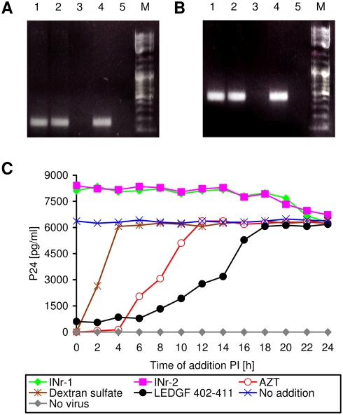
Figure 8. Virus-cell adsorption and viral reverse-transcriptase activity are not affected by the INr peptides.
(A and B) Estimation of viral DNA in virus-infected cells: Sup-T1 cells were incubated with the indicated peptides or with AZT for 2 h and then were infected with HIV. Following another 6 h incubation, the viral Gag (A) or Nef (B) DNA sequences were amplified using specific primers. The reaction was terminated after 30 cycles. Lanes: 1) INr-1 (12.5 µM); 2) INr-2 (12.5 µM); 3) AZT (2 µM); 4) untreated cells; 5) uninfected cells. Note that in the AZT-treated cells, reverse-transcribed viral DNA is absent. (C) Influence of the time of addition on P24 production [59]: Sup-T1 cells were infected with HIV-1 at a MOI of 2, and the indicated constituents were added at different time points PI (0, 2, 4,…, 24 h). Viral p24 was determined at 48 h PI. No addition (Blue X); no virus (Gray diamond); dextran sulfate 20 µM (Brown asterisk); AZT 2 µM (Red empty circle); INr-1 12.5 µM (Green diamond); INr-2 12.5 µM (Magenta square); LEDGF 402–411 (Black full circle). All other experimental conditions are described in Materials and Methods.

