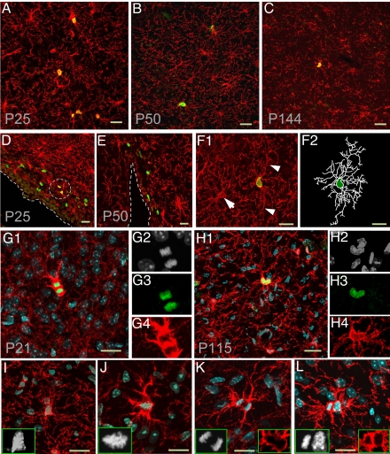
Fig. 1.
Dividing NG2 cells with differentiated morphology in adult brains. (A–C) Immunostaining with antibodies against NG2 (red) and BrdU (green) in cerebral cortex from P25 (A), P50 (B), and P144 (C) mice. (D and E) Immunostaining with antibodies against NG2 (red) and BrdU (green) in SVZ region from P25 (D) and P50 (E) mouse brains. NG2+BrdU+ cells are circled. (F) Example micrograph showing the differentiated morphology of 1 NG2+BrdU+ cell from a P50 mouse brain 2 h after BrdU pulse-labeling. Arrowheads point at 2 long processes of the BrdU+ cell, and the arrow points at a BrdU− cells. The processes and cell body of the dividing NG2 cell were outlined with soft Neurolucida (F2). (G and H) Immunostaining with antibodies against NG2 (red) and Ki67 (green) in the cortex of P21 (G1–G4) and P115 (H1–H4) mouse brain, respectively. Nuclei of dividing cells were strongly labeled by DAPI (white, G2 and H2) and Ki67 (G3 and H3). The Ki67+ cells in telophase (G1) and prophase (H1) both bear multiple processes. (I--L) Four dividing NG2 cells (red) labeled by DAPI (white) and anti-NG2 (red) in different mitotic stages: prometaphase (I), metaphase (J), anaphase (K), and telophase (L). Note the difference between the shape and density of DAPI signal in dividing cells and nondividing cells (I–L). Note NG2 expression was up-regulated during mitosis (G, J, and K). All scale bars are 20 μm.

