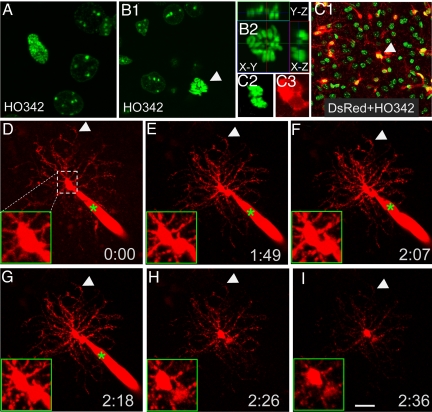
Fig. 2.
Monitering NG2 cell division while retaining differentiated morphology. (A and B) The dividing cells in prophase (A) and metaphase (B1 and B2) labeled by HO342 in acute brain slices. (B2) 9.6 μm Z stack with orthogonal views at the point of dividing nucleus. (C1–C3) A dividing cell of telophase (C2) in an acute slice from a P13 NG2DsRedBac transgenic mouse. (C2) HO342. (C3) DsRed. (B and C) Arrows point at the dividing cells. (D–I) Time-lapse recording of a dividing cell (red) filled with Alexa Fluor 568 from recording pipette (green star). Note the long multiple processes that remained unretracted during dividing. (D–I) Arrows indicate 1 process remaining at the same place during division. (D–I, Insets) Higher magnification views of the somatic region of the cell.(Scale bar, 20 μm.)

