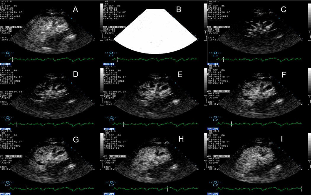
Figure 2.

Assessment of renal blood flow using CEU. Panel A, steady state; Panel B, destruction of microbubbles in the tissue using high energy ultrasound. Panels C through I, replenishment of the tissue with microbubbles. Note, almost immediate appearance of microbubbles in the main arteries followed by cortex and finally the medulla.
