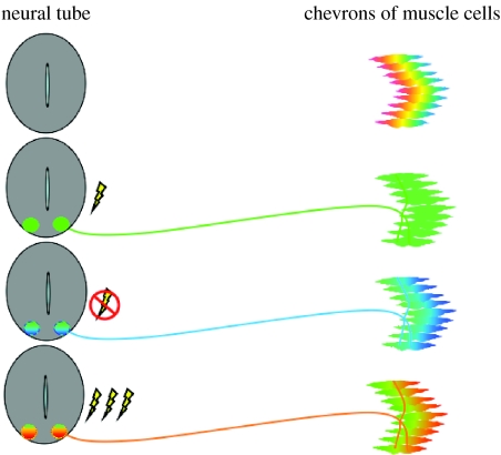
Figure 4.
Model illustrating the process of transmitter–receptor matching at the NMJ. Prior to innervation, muscle cells express multiple classes of receptors. During normal development, motor neurons express ACh and AChR that persist on the muscle fibres. When spontaneous embryonic calcium spike activity is blocked, motor neurons express glutamate in addition to ACh, and both AChR and GluR are found on muscle fibres. When calcium spike activity is increased, motor neurons express GABA and glycine and the cognate receptors are found on muscle fibres. Colour coding of transmitter receptors indicates their presence but not their distribution on the muscle surface (red: GABAR, GABA; yellow: glyR, gly; green: AChR, ACh; blue: gluR, glu.).

