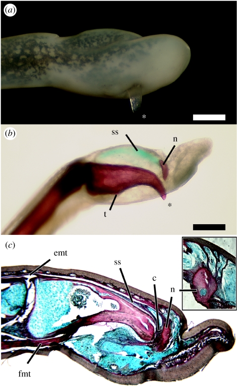
Figure 1.
Anatomy of the claws of Astylosternus and Trichobatrachus. (a) Right fourth toe of Astylosternus rheophilus (MCZ A-136934), in lateral view. The exposed bony tip of the terminal phalanx (asterisk) protrudes through the skin on the ventral side. (b) Cleared and stained left fourth toe of Astylosternus laurenti (CAS 158971) in medial view. Bone is red; the suspensory sheath is stained blue. The tendon of the deep digital flexor muscle inserts on a tubercle (t) of the ventral surface of the phalanx. The bony nodule (n) is connected to the proximodorsal surface of the terminal phalanx by a suspensory sheath (ss). (c) Longitudinal section through the left fourth toe of Trichobatrachus robustus (MCZ A-137591) showing the claw in resting state. The terminal phalanx is linked to the bony nodule (n) via collagen-rich tissue (c, red) and is underlain by dense, collagen-poor connective tissue (blue). Activation of the deep digital flexor tendon (fmt) breaks the phalanx free from the bony nodule and exposes the bony tip. The suspensory sheath (ss) connecting the bony nodule to the proximodorsal phalanx shares a common insertion with the tendon of the digital extensor muscle (emt). Inset: longitudinal section through right third toe of T. robustus (MCZ A-137951) with an exposed claw, stained as in c; close-up of the bony nodule. The nodule remains anchored to the dermis via collagenous strands (red). Scale bar, 0.5 mm.

