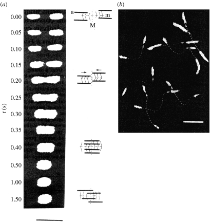
Figure 2.
In vitro motility of fluorescently labelled actin filament. (a) Sequence fluorescence micrographs of a sarcomere containing fluorescently labelled actin filament without Z-lines during shortening. Scale bar, 5 μm. (b) Sliding movement of fluorescently labelled actin filaments on a glass slide coated with single-headed myosin filaments. Two images taken 1.5 s apart were photographed on the same frame. Dotted lines represent traces of the movement of actin filaments with arrowheads indicating the direction of the movement. Scale bar, 5 μm.

