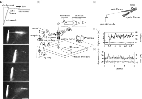
Figure 3.
Manipulation of single actin filaments and force measurement using a microneedle. (a) Measurement of the tensile strength of an actin filament. A single actin filament bound to a flexible glass microneedle at one end (at the left) was stretched through a stiff needle bound at the other end until the actin filament was broken. The flexible needle was bent as the filament was stretched. (b–d) Force measurement of the interaction between myosin molecules and actin filament. (b) Schematic of the measurement system with nanometre-, piconewton- and millisecond-order sensitivity. See Ishijima et al. (1996) for details. (c) A single actin filament was captured by a glass needle and interacted with small number of myosin heads in the cofilament of myosin and myosin rod. (d) The force fluctuation exerted by individual myosin molecules was measured as a displacement record of a tip of a microneedle. (e) Displacement record caused by single myosin heads. The lower trace indicates thermal vibrations of free needles. Broken lines show ±5 nm.

