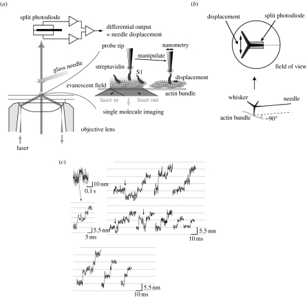
Figure 7.
Manipulation of single myosin molecules using a scanning probe and measurement of substeps of myosin. (a) Schematic of the measurement. See reference Kitamura et al. (2005) for details. (b) The schematic of the tip of the scanning probe. A single myosin head was captured at the tip of a whisker attached at the glass microneedle. Motion of myosin head is restricted geometrically. (c) Stepwise movements in the rising phase of the displacements. Some backward steps were observed as indicated by arrows.

