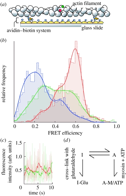
Figure 8.
Single molecule FRET measurement from single actin molecules in the filament. (a) Schematic of the experiment. Actin monomer in the filament was specifically labelled with tetramethyl-rhodamine (TMR) attached as a donor and IC5 attached as an acceptor. (b) Histograms of FRET efficiency for actin molecules. Blue bars and line, actin molecules cross-linked with glutaraldehyde. Red bars and line, actin molecules in the presence of myosin V. Green bars and line, intact actin filaments. (c) Time course of the donor (green line) and the acceptor (red line) fluorescence from a double-labelled actin. (d) The conformational dynamics of actin molecules in the actin filament.

